Child
- About
- Meet The Team
- Conditions
- Aortic Stenosis
- Atrial Septal Defect
- Coarctation of the Aorta
- Complete Atrioventricular Septal Defect
- Heart Murmur
- Interrupted Aortic Arch
- Normal Heart
- Partial Atrio-Ventricular Septal Defect
- Patent Ductus Arteriosus
- Pulmonary Atresia with Intact Ventricular Septum
- Pulmonary Atresia with Ventricular Septal Defect
- Pulmonary Stenosis
- Right Aortic Arch
- Small Ventricular Septal Defect (Muscular)
- Small Ventricular Septal Defect (Perimembraneous)
- Tetralogy of Fallot
- Transposition of the Great Arteries
- Ventricular Septal Defect (Large)
- Dental Practitioners: Dental care in children at risk of Infective Endocarditis
- Looking after your child’s oral health
- Coming for an echocardiogram
- Outpatient Appointments
- Preparing to Come into Hospital for Surgery
- On Admission to the Children's Ward
- Visiting
- Operation Day
- Children's Intensive Care
- Daily Routine on Intensive Care
- Managing your child's discomfort
- Going Home
- Children's Cardiac MRI Scan
- Cardiac Catheter
- Reveal Device
- Ablation Procedure
- Pacemakers
- INR and Warfarin
- Lifestyle and Exercise Advice
- School Advice
- Attachment
- Yorkshire Regional Genetic Service
- Advice & Support Groups
- Your Views
- Monitoring of Results
- Second Opinion
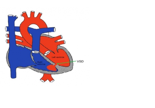
Small Ventricular Septal Defect (Muscular)
A muscular ventricular septal defect is a hole in the muscle wall between the two main pumping chambers of the heart (the ventricles). In the normal heart the left ventricle works at high pressure and pumps blood to the body and the right ventricle works at low pressure and pumps blood to the lungs. When
there is a hole between the two ventricles (a VSD), blood flows from the left ventricle to the right ventricle through the hole. Some patients may have more than one VSD.
Most holes in the thick muscular part of the ventricular septum gradually get smaller or even close off completely on their own as the child grows. In some cases this happens within a few months, in others it may take many years and in some the VSD doesn’t close at all. Even if a small VSD does not close by itself, it does not usually need any treatment and does not usually stop the patient leading a completely normal life.
Tests
An ultrasound scan of the heart (“an echocardiogram”) is required to measure the size of the VSD is and see how many VSDs there are.
General advice for the future
Patients with small VSDs can live normal active lives, including all kinds of sport.
All patients with a VSD will be at risk of infection in the heart (called endocarditis). Such infections may be caused by infections of the teeth or gums. It is important to look after your child’s teeth and visit the dentist regularly. Ear or body piercing and tattooing are best avoided as they also carry a small risk of infection which may spread to the heart. If a VSD closes by itself these precautions are no longer necessary.