Adult
- About
- Meet The Team
- Conditions
- Anticoagulation in Pregnancy
- Aortic Dilatation and Pregnancy
- Aortic Valve Disease
- Aortic Valve Replacement
- Atrial Septal Defect
- Coarctation - Transcatheter stent (keyhole) treatment
- Coarctation of the Aorta
- Congenitally Corrected Transposition of the Great Arteries
- Ebsteins Anomaly
- Eisenmenger’s Syndrome
- Fontan Circulation
- Mitral Valve Repair/Replacement
- Normal Heart
- Patent Foramen Ovale
- Pregnancy information for women with metal heart valves
- Pulmonary Incompetence
- Pulmonary Stenosis
- Pulmonary Valve Replacement - Surgery
- Pulmonary valve replacement - Transcatheter (keyhole) treatment
- Repaired Atrioventricular Septal Defects
- Sub-aortic Stenosis
- Surgical treatment of Atrial Septal Defect
- Tetralogy of Fallot
- Transposition of the Great Arteries - The Atrial Switch (Mustard or Senning) procedure
- Transposition of the Great Arteries – Arterial Switch
- Ventricular Septal Defect
- Ventricular Septal Defect - Transcatheter (keyhole) treatment
- Patient Feedback
- Making the most of your clinic appointment
- Your Appointment in Outpatients
- Easy Read Guide for Out Patients
- Cardiac Catheter
- Transoesophageal Echocardiogram
- MRI
- Surgery & "Top Tips" for coming into hospital
- Lifestyle Advice
- Exercise
- Heart Failure
- End of Life and Palliative Care
- Looking after your oral health
- Dentists Information Section: Dental care in adults at risk of Infective Endocarditis
- Yorkshire Regional Genetic Service
- Support
- Video Diaries
- Second Opinion
- Monitoring Results at Leeds Infirmary
- Professionals
Pulmonary Valve Replacement - Surgery
The pulmonary valve is located between the right-sided pumping chamber of the heart and the lung arteries. Blood from the body comes into the right side of the heart and is then pumped to the lungs where it picks up oxygen. The blood from the lungs returns to the left side of the heart and is then pumped back to the body. The function of the pulmonary valve is to ensure that blood only travels from the right side of the heart chamber to the lung arteries and does not leak back the other way.

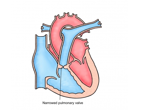
In some cases, the leaflets of the pulmonary valve can be abnormal. Valve leaflets can be thickened in which case the medical term for the problem is pulmonary stenosis. If the valve leaks then the medical term for this is pulmonary regurgitation. Leakage of the valve is particularly common in those patients who have had surgery for “Tetralogy of Fallot” during childhood. Both narrowing and leak can result in the muscle of the right-sided pumping chamber (right ventricle) having to work harder than normal. The muscle can become thickened or the chamber can stretch to a size that is bigger than usual. In most cases this is not a serious problem and needs no treatment. However, if the narrowing or leaking is severe then the heart cannot pump normally and an operation to replace the valve can be necessary. The diagram above shows a normal heart on the left and a heart with a narrowed pulmonary valve on the right.
Tests
The Operation
Pulmonary valve problems can be treated by an open heart operation where the valve is replaced. The operation is performed under a general anaesthetic so you will be asleep. The surgeon makes an opening in the middle of the chest through the breast bone to get to the heart. During the operation, the function of the heart and lungs is taken over by a machine (heart bypass). The surgeon can then stop the heart and remove the damaged valve. The new replacement valve can then be sewn in place. After the operation the breastbone is closed using stainless steel wires and surgical drains (tubes) are positioned in the chest to allow any excess fluid to drain. These are normally taken out after about 24 hours. As a precaution, pacing wires are placed on the surface of the heart in case of a slow heart rate in the early post-operative stage; these are removed 4-5 days after the operation.
There are 2 types of valve which are commonly used:
Tissue Pulmonary Valves – engineered from a pig or cow heart valve.
Homograft Valves – from a donor human heart.
There is a very small risk of death (less than 1 in 100) and a very small risk of major complications such as brain damage (less than 1 in 100). Other complications such as bleeding, infection, fluid collecting around the heart or lungs can occur after the operation but these are rarely serious, although they may need treatment. After surgery a short stay on the intensive care unit (usually 2 days) is required. You will then remain in hospital for further monitoring. This is usually for about 5 days but can be longer. Opening the front of the chest leads to a scar and the chest wall will be sore whilst it heals. The time taken to get fully back to normal varies from person to person but can be up to 3 months.
Tests
After the Operation
After your operation you will be closely monitored by both the surgeon and cardiologist. A nurse specialist will visit you on the ward and give you advice about the period after discharge and answer any questions that you may have. A nurse will phone you at home in the week following discharge to check on your progress. You will be given a telephone number/email address that you can contact should you have any concerns in between discharge and follow-up
Tests
Final Points
In the 2 weeks leading up to your surgery, you will receive an appointment for the preadmission clinic. This is to make sure that you are fit for surgery and also to give you an opportunity to find out more about coming into hospital and ask any questions.
It is also important to make sure that all dental work has been completed within 6 months before your surgery to reduce the risk of infection in the heart (endocarditis).
You will need to take some medication following the operation but it will most likely be temporary.
You will need between 6 and 12 weeks off work, depending on your job.
You cannot drive for 6 weeks following the operation.
